DW-F3 Trolley Color Doppler Ultrasound: Features, Uses, and Benefits
Discover the DW-F3 Trolley Color Doppler Ultrasound System: portable, high-resolution imaging with advanced blood flow analysis for versatile clinical diagnostics.
Introduction to Ultrasound and Doppler Imaging
Understanding Ultrasound Imaging
Sonography or ultrasound imaging involves the use of sound waves of high frequency to visualize the internal tissues, organs, and flow of blood. It is non-invasive, radiation-free free and real-time imaging, making it perfect for the diagnostics of cardiology, obstetrics, gynecology, and general medicine.
Color Doppler Technology
Color Doppler is an imaging technique that provides blood flow analysis to standard grayscale imaging. It imagines the flow direction and speed as color overlays to assist clinicians in assessing the integrity of the vessels, identifying obstacles, and evaluating heart performance. The DW-F3 combines color Doppler and several imaging modes that boost diagnostic accuracy.
Define DW-F3 Trolley Color Doppler Ultrasound
The DW-F3 Trolley Color Doppler Ultrasound Scanner System is an ultra-high-performance, trolley-based diagnostic imaging system that can be used in a variety of hospital, clinic, and imaging facilities.
The DW-F3, based on a complex of high-resolution imaging, advanced color Doppler, and a mobile, ergonomic design, supports effective diagnostics in a variety of clinical types of activity.
What is Color Doppler Ultrasound?
Color Doppler is an ultrasound mode that demonstrates blood circulation within vessels by way of color.
- The direction and speed of the blood flow are indicated by red or blue colors.
- Assist physicians in identifying abnormal heart conditions, clots, or blood circulation problems.
- Usually conducted during pregnancy to monitor the blood flow and a healthy heart of the baby.
This is what differentiates the DW-F3 as more developed compared to the plain ultrasound, which only displays the organs in grayscale.
Key Features of DW-F3 Trolley Color Doppler Ultrasound System
1. Mobile Trolley Design
The trolley on the DW-F3 is also on wheels, making it easy to move across rooms without compromising on stability. Its small size simplifies the workflow and is therefore suitable in a hospital that needs scanning options among various departments.
2. Comprehensive Color Doppler Imaging
The system has a color Doppler, pulse wave Doppler, linear, convex, and transvaginal probes with which blood flow and internal organs can be fully visualized and used in various clinical applications in different specialties.
3. High-Resolution Imaging
High contrast noise-reduced images are made possible by advanced technologies such as tissue harmonic imaging and spatial compounding. This increases the level of diagnostic accuracy as clinicians can see the tissues, vessels, and organs at the highest levels.
4. Wide Range of Probe Compatibility
Supports convex (2-5.5 MHz), linear (6-12 MHz), and transvaginal (5-9 MHz) probes and is versatile in imaging the abdomen, superficial, gynecological, and musculoskeletal examinations in clinical environments with many different departments.
5. User-Friendly Interface
Large LCD screen, intuitive control panel, pre-established imaging settings, and automatic optimization in a one-touch feature make scanning easier, help lessen the learning curve, and make scanning more productive when the clinic is busy.
6. Data Management and Storage
Built-in memory and USB will enable the storage, retrieval, and sharing of patient images safely. Clinicians can efficiently handle high volumes of data in the documentation, consultation, and record-keeping.
Clinical Applications of the DW‑F3 Scanner
Obstetrics and Gynecology
The DW-F3 is used to track fetal development, evaluate the health of the placenta, and also test reproductive organs. Color Doppler enables one to see the fetal heart rate and umbilical blood circulation, which is useful in prenatal care.
Cardiology
The system is able to identify abnormal blood flow, valve complications, and abnormalities in the heart chambers, which assists in diagnosing cardiovascular diseases.
Abdominal Imaging
It gives detailed pictures of organs such as the liver, kidney, bladder, and spleen, which are used in the identification of tumors, cysts, stones, or structural abnormalities.
Musculoskeletal and Small Organ Scans
Linear probes allow high-resolution imaging of the tendons, ligaments, and superficial organs, which are necessary in orthopedic or sports medicine.
Pediatrics and Neonatal Care
It is also applicable in imaging babies and newborns because its sensitive probes and intense imaging are important in cases where accuracy matters.
Why Choose DW-F3?
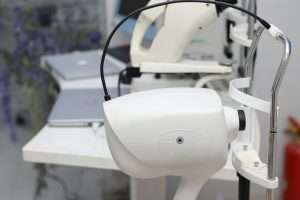
The DW-F3 Trolley Color Doppler Ultrasound Scanner System is portable, has high-quality imaging, and multi-probe versatility, which makes it a good fit in hospitals, clinics, and diagnostic centers that have to find good solutions for diagnostic ultrasound. Its trolley construction gives it efficiency in the workflow with high-resolution color Doppler imaging and a vast clinical application.
Pricing and Availability
The DW-F3 system cost can be purchased between 4,500 and 337,500 by Acute Pharmacy, depending on configuration and the choice of probe. Bulk buying of several units is possible, and it can be customized or not, depending on the needs of the hospital.
Who Can Use It?
The DW‑F3 is suitable for:
- General practitioners and radiologists.
- OB/GYN and prenatal clinics
- Urology and vascular imaging unit.
- Emergency and inpatient care.
- Privately owned healthcare facilities and hospitals.
Its flexibility enables it to be a wide-ranging instrument for both the preliminary examination and exact follow-up analysis.
Workflow and Software Features
User-Friendly Interface
- Backlit keys, ergonomic control panel.
- Ready-made pre-scanning settings.
- Single-click optimization of uniform image quality.
Image Storage and Connectivity
- External USB saving and internal storage.
- PACS integration compatibility with DICOM 3.0.
- Convenient access and transmission of pictures
Multi-Mode Functionality
- Quick alternation of 2D, color Doppl, er and superior imaging specifications.
- Allows support of imaging and measurement processes at the same time
Tips for Healthcare Facilities
Training and Operation
The practical training of staff is to be provided so that they can take advantage of Doppler modes successfully, particularly in cases of vascular studies.
Probe Preservation
Adequate cleaning and storage of probes will guarantee the maintenance of the same performance and increase the longevity.
Integration with PACS
The ultrasound system should be combined with the PACS (Picture Archiving and Communication System) to facilitate the management of the records in the hospitals.
FAQs
Is the DW‑F3 portable?
Yes. It is attached to a stable, movable trolley, which makes it easy to move around the rooms and bedside examinations, but remains stable during scanning.
Can the DW‑F3 be integrated with hospital systems?
Yes. It aids PACS (Picture Archiving and Communication System) integration, which enables the effortless storage, retrieval, and exchange of images and reports of patients.
Does the DW‑F3 support multi-language operation?
Yes. It also has more than one language, which is both English and French, so it can be used in an international clinical setting.
Conclusion
DW-F3 Trolley Color Doppler Ultrasound Scanner is a versatile probe that integrates mobile capability, high-resolution imaging, and versatility in the high-end positioning of its product in the market by catering to hospitals, clinics, and diagnostic centers. Its high image color Doppler technology improves blood flow examination, which guarantees correct, effective, and dependable diagnostics in various clinical uses.